
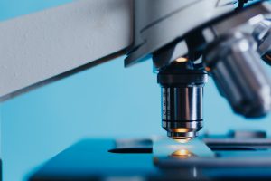
Cancer is the second leading cause of death in the world.1 In 2018, an estimated 17 million people worldwide were newly diagnosed with cancer, an increase from 12.3 million in 2007. When a doctor suspects cancer, the first step in the diagnosis process is usually a biopsy, a microscopic examination of a patient’s tissue sample performed by a pathologist. After the sample is prepped, the tissue undergoes hematoxylin and eosin staining. This process stains different cells and structures so that they’re visible under a microscope. The pathologist can then detect any abnormalities. In most cases, this is the only step needed for diagnosis.
In other cases, specialized staining is needed to determine amounts of specific proteins or the presence of certain genes, which then determines how a patient might respond to treatment. In an immunohistochemistry (IHS) test a diagnostic antibody is used to detect proteins in cells or cell membranes. If the specific protein looked for is present, the antibody binds to it. This can be detected as a color change under a microscope. In this way, an IHS test can determine what type of drug therapy will be most effective. An IHS test can also detect if a tumor is benign or malignant.
Beyond cancer diagnosis, IHS tests are used to diagnose autoimmune, cardiovascular, infectious, and nephrological diseases. They are also used to diagnose diabetes. In medical research laboratories, they’re used to test the effectiveness of new therapeutic drugs. This type of test is considered the most profitable tissue diagnostic test. In 2017, IHS test revenue was valued at $1.6 billion.
Other types of tissue diagnostic tests include fluorescent in situ hybridization and silver in situ hybridization. They are used to detect the presence of human epidural growth factor receptor 2 proteins, also known as HER2 proteins, on breast cancer tumors. Tumors that tend to grow and spread faster have higher than normal levels of the HER2 protein. These tumors respond better to newer cancer drugs that specifically target HER2-positive tumors.
Today’s market size shows the total revenue from tissue diagnostic tests globally in 2018 and projected for 2025. Demand for tissue diagnostics is expected to grow due to an aging population and rising incidence of chronic diseases worldwide. Not surprisingly, hospitals claimed half of the market share due to the high volume of tests done for disease diagnosis, monitoring, and treatment. Research laboratories claimed about 25% of the market. Pharmaceutical organizations and clinical research organizations combined claimed the rest. Leading companies in this industry include F. Hoffmann-La Roche Ltd., Abbott, Siemens Healthineers, and QIAGEN.
1 According to the World Health Organization, cardiovascular diseases claimed 17 million, cancer 9 million, and infectious and parasitic diseases 5.5 million lives worldwide in 2018.Geographic reference: World
Year: 2018 and 2025
Market size: $3.9 billion and $6.03 billion, respectively
Sources: “Tissue Diagnostics Market Analysis Report by Technology (Immunohistochemistry, In Situ Hybridization, Digital & Anatomic Pathology), by Application, by End Use, and Segment Forecasts, 2019 – 2025,” Grand View Research Report Summary, January 2019 available online here; “Tissue Diagnostics Market Worth $6.03 Billion by 2025 | CAGR: 6.6%,” Grand View Research Press Release, January 2019 available online here; Global Cancer Facts & Figures 4th Edition, American Cancer Society, 2018 available online here; Global Cancer Facts & Figures 2007, American Cancer Society, 2007 available online here; “What Happens With a Biopsy?” F. Hoffman-La Roche Ltd., 2019 available online here; “Breast Cancer HER2 Status,” American Cancer Society, September 25, 2017 available online here.
Image source: Konstantin Kolosov, “analysis-biochemistry-biologist-2030265,” Pixabay, February 1, 2017 available online here.